
Figure 1.
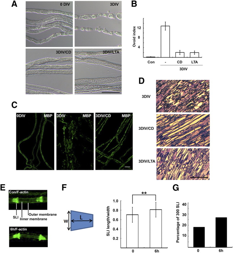
Actin polymerization is required for WD in vitro. A, Sciatic nerve explants were cultured for 3 d (3 DIV) in the absence or presence of CD (5 μm), and then the explants were fixed, and teased nerve fibers were prepared. B, Quantitative result of the number of myelin ovoids. C, Teased nerve fibers were immunostained with an antibody to MBP. D, Sciatic nerve explants were fixed and processed for semithin section analysis. Scale bar, 50 μm. E, Enlarged images showing the distribution of F-actin in the SLI and Schwann cell membranes. The nonlabeled area between inner and outer membranes is the compact myelin sheath. F, The width (W) and length (L) of F-actin-stained SLI were measured, and the index indicates length/width. **p < 0.01, Student's t test. Error bar indicates SD of 300 SLIs from three different experiments. G, The percentage of SLIs from the control (Con) and 6 h culture groups with the length/width indices >1 (N = 300 SLIs per group).