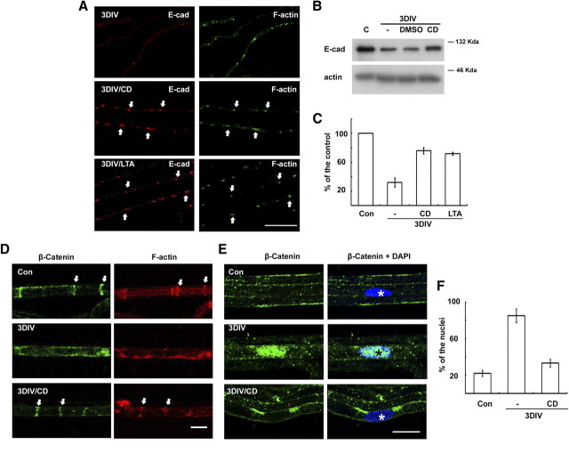
Figure 3.
Actin polymerization-dependent regulation of E-cadherin and β-catenin in the SLI during WD. A, Teased nerve fibers were immunolabeled with an antibody against E-cadherin and fluorescein phalloidin. Arrows indicate the SLI where E-cadherin and F-actin are colocalized. Scale bar, 100 μm. B, Protein extracts (10 μg) from sciatic nerve explants were analyzed by Western blotting. Dimethylsulfoxide (DMSO) was the vehicle for drugs. C, Quantitative analysis of Western blotting for E-cadherin expression from three independent experiments. D, E, Immunofluorescent staining against β-catenin in the teased nerve fibers from in vitro cultures. Arrows in D indicate SLI, whereas asterisks in E indicate the nucleus. Scale bar, 20 μm. F, Quantitative analysis of the nuclear immunofluorescent staining against β-catenin from three independent experiments. The percentage of β-catenin-reactive nuclei from DAPI (diamidino-2-phenylindole dihydrochloride)-positive nuclei (∼100 nuclei from each experiment) in the teased fibers was calculated. C and Con, Control; E-cad, E-cadherin.

