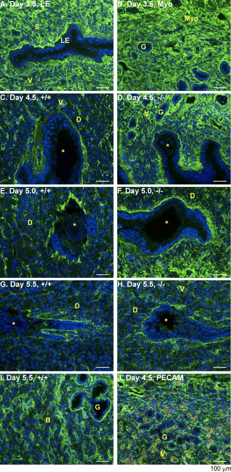
FIG. 4.
Spatiotemporal expression of COL VI in the WT and Lpar3−/− uterus. Fresh frozen uterine cross sections (Day 3.5 and Day 5.5) and longitudinal sections (Day 4.5 and Day 5.0) were cut at 10 μm. A) Day 3.5 uterus to highlight LE and stroma. B) Day 3.5 uterus to highlight GE and myometrium; there is no obvious difference in the COL VI labeling between Day 3.5 WT and Lpar3−/− uterus. C) WT, Day 4.5. D) Lpar3−/−, Day 4.5. E) WT, Day 5.0. F) Lpar3−/−, Day 5.0. G) WT, Day 5.5. H) Lpar3−/−, Day 5.5. Green color represents COL VI, blue represents DAPI, red represents PECAM, and the yellow star indicates the embryo. B, basal endometrium; D, decidual zone; G, glandular epithelium; Myo, myometrium; S, stroma; V, vasculature. Bar = 100 μm; n = 2–3.

