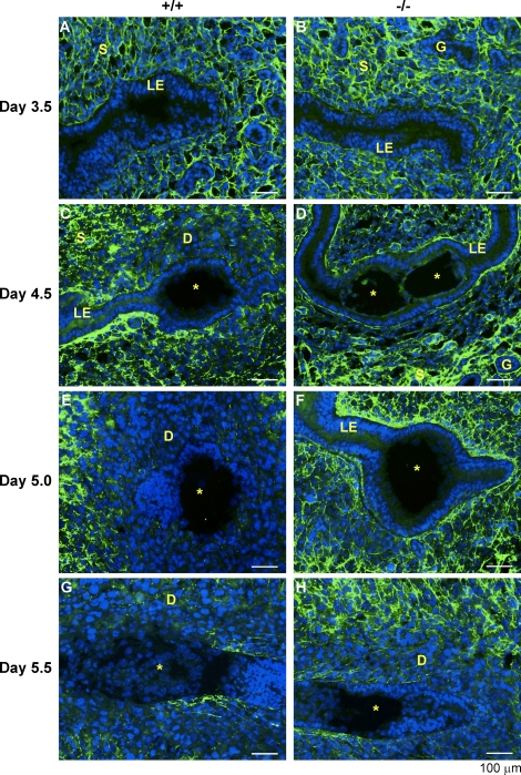
FIG. 5.
Distribution of COL I immunoreactivity in the WT and Lpar3−/− uterus from Day 3.5 to Day 5.5. Fresh-frozen uterine cross sections (Days 3.5 and 5.5) and longitudinal sections (Days 4.5 and 5.0) were cut at 10 μm. A) WT, Day 3.5. B) Lpar3−/−, Day 3.5. C) WT, Day 4.5. D) Lpar3−/−, Day 4.5. E) WT, Day 5.0. F) Lpar3−/−, Day 5.0. G) WT, Day 5.5. H) Lpar3−/−, Day 5.5. Blue color indicates DAPI, green represents COL I, and the yellow star indicates the embryo. D, decidual zone; G, glandular epithelium; LE, luminal epithelium; S, stroma. Bar = 100 μm; n = 2–3.

