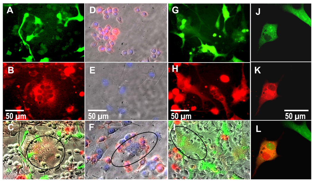
Figure 3.
Fluorescence images showing A) RAW macrophages labeled green, B) 3T3 fibroblasts labeled red, and C) an overlay of brightfield and red and green channels showing a red (circled), and therefore fibroblastic, multinucleated cell. Fluorescence images showing D) macrophages and E) fibroblasts incubated with DAPI (blue) and also fluorescently labeled anti-CD14 (red), revealing F) fibroblasts that stain negative for CD14, are multinucleated (circled), and are therefore not macrophages. Fluorescent G–I) and confocal J–K) images revealing co-localization of a red-labeled population of fibroblasts fused with a green-labeled population of fibroblasts in the presence of non-labeled macrophages, where G and J are the green channel, H and K are the red channel, and I and L are overlays of I) brightfield and red and green fluorescent channels and L) red and green channels (co-localization experiments with 3 replicates each were repeated 3 times). Images shown after 24 hours of culture.

