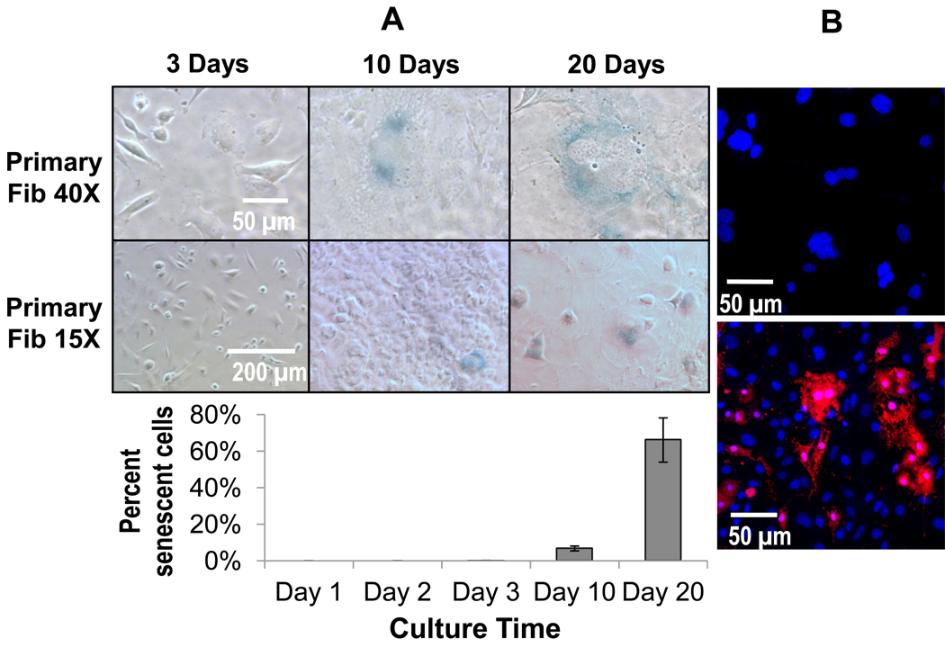
Figure 8.
Panel A) Senescence staining (blue) in primary fibroblasts after 3, 10, and 20 days expressed qualitatively as representative images and quantitatively in graph below. These data show that at longer time points near day 10, senescence becomes prevalent in primary fibroblasts. Data are represented as the mean ± SEM from 3 independent replicates. Importantly, all multinucleated fibroblasts regardless of time point, stained positive for senescence. Panel B) Confocal images showing DAPI (blue) and apoptosis staining (red) in primary fibroblasts (top image) left untreated and (bottom image) treated with bupivicaine as a positive control. Multinucleate fibroblastic cells even with highly polymorphic nuclei did not stain positive for apoptosis. Images shown after 3 days in culture.

