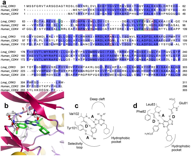
Figure 2. Predicted binding of azapurine pharmacophore and model of CRK3 active site.
a) Sequence alignment of LmajCRK3, Human CDK2 and Human CDK4 showing the percentage identity in shades of blue. The active site regions are boxed in green with key differences boxed in red. b) A model of Lm CRK3 with an azapurine derivative (compound 11) docked in to the ATP site to show the predicted binding mode. c) Schematic overview of the predicted binding mode of azapurine derivatives with Lm CRK3 detailing the A-D-A motif not possible in CDK2 due to the Tyr101 - Phe82 difference. d) CDK2 binding mode with NU6102 [39] showing the D-A-D motif.

