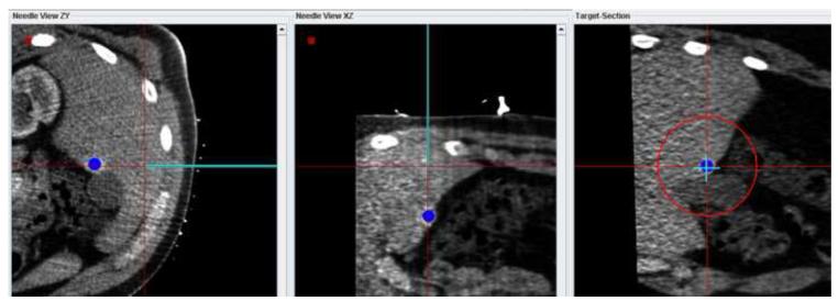
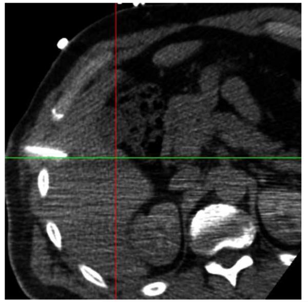
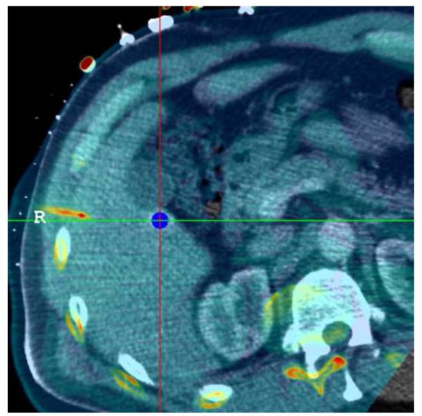
Figure 6.
(a) Navigation display showing the virtual needle aligned with the target identified in the arterial phase-enhanced CT. (b) Non-contrast enhanced confirmation scan of the needle position does not show the target lesion. (c) After rigid registration of the confirmation scan (pseudo-colored) with the arterial contrast-enhanced scan (gray scale background) in the vicinity of the target lesion, the correct alignment of the needle with the target is confirmed.


