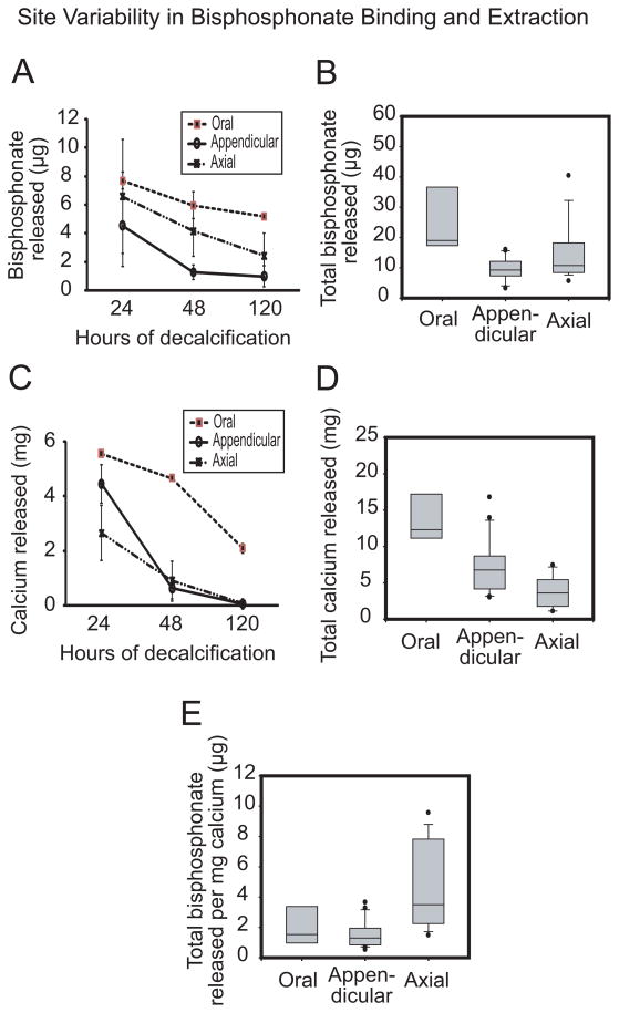
Figure 3.
Regional variability in skeletal uptake and release of fluorescently labeled bisphosphonate. Time-dependent release of hydroxyapatite-bound bisphosphonate (A) and calcium (C) as well as total bisphosphonate (B) and calcium (D) released were significantly highest in oral bones relative to axial and appendicular sites (p < 0.05); an indication that bisphosphonate can be readily liberated from the mandible due to the ease of calcium release. Bisphosphonate per unit calcium (E) was highest in axial bone relative to oral and appendicular bones [n = 4; median is the line within the box; the ends of the boxes define 25th and 75th percentiles; the two bars outside the box define 10th and 90th percentiles].

