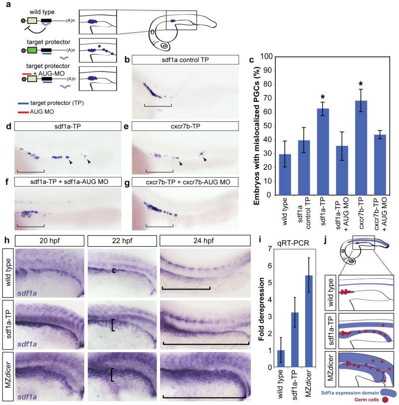
Figure 3.
Blocking miR-430-mediated repression of sdf1a and cxcr7b causes PGC mislocalization and expanded sdf1a expression. (a) Schematic representation of the experimental setup. Injecting the TP (purple) blocks miRNA-mediated repression, increasing mRNA expression and leading to mislocalization of cells. Co-injecting a morpholino to reduce translation of the target gene (red, AUG MO) rescues the mislocalization phenotype. The inset shows the region of the embryo depicted in the panels (b, d-h). (b, d-g) Whole mount in situ of nanos mRNA, labeling PGCs in 24 hpf embryos. Bracket shows correct localization of PGCs. Arrowheads identify mislocalized PGCs. (c) Quantification of the percentage of embryos with mislocalized PGCs in each experimental condition as indicated. A significantly increased number of TP-injected embryos have mislocalized PGCs (*, p=1.185 ×10⁻⁷, sdf1a-TP; p=2.52×10⁻⁷, cxcr7b-TP; two-sided Fisher’s exact test). Error bars show ± s.d. (d, e) Representative images of PGC mislocalization are shown. (f, g) Co-injection of a low level of the corresponding AUG MO rescues the TP phenotype (sdf1a AUG MO, 0.01 pmol; cxcr7b AUG MO, 0.045 pmol). (h) In situ hybridization to detect sdf1a mRNA. The trunk of embryos at 20 hpf, 22 hpf, and 24 hpf are wild type, injected with sdf1a-TP, or MZdicer. Brackets illustrate the extension of the sdf1a expression domain along the pronephric region. (i) qPCR for sdf1a in 24 hpf wild type, sdf1a-TP-injected embryos, and MZdicer. An increase in expression was observed in the absence of miR-430-mediated repression. (j) Schematic summary of Sdf1a tail expression and the resulting PGC mislocalization.

