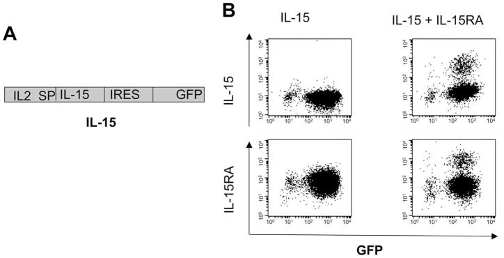
FIGURE 1.
Characterization of the expression of IL-15 on TC-1 cells transduced with IL-15 and/or IL-15RA. A, Schematic diagram of the IL-15 construct. TC-1 cells were transduced with a retrovirus encoding the IL2SP linked to IL-15 followed by IRES and GFP. This construct is denoted as IL-15. The IL-15-transduced TC-1 cells were sorted for GFP expression, and a portion of GFP+ cells was further transduced with retrovirus-encoding IL-15RA. Cells were analyzed for IL-15 or IL-15RA expression by flow cytometry analysis. B, Representative flow cytometry data demonstrating the expression of IL-15 and IL-15RA in cells transduced with IL-15 and/or IL-15RA. Note that surface expression of IL-15 was significantly higher on the TC-1 cells transduced with both IL-15 and IL-15RA. The data shown here are from one representative experiment of two performed.

