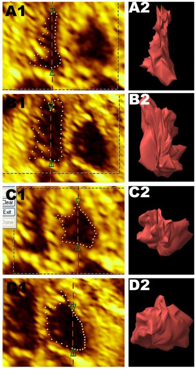
Figure 1.
Selected VOCAL rotational steps utilizing Contour Finder: Trace for each ventricle in end systole and end diastole (A: Left ventricle in systole; B: Left ventricle in diastole; C: Right ventricle in systole; D: Right ventricle in diastole) at the level of the four chamber view (A1-D1) and the rendered image (A2-D2).

