
Figure 3.
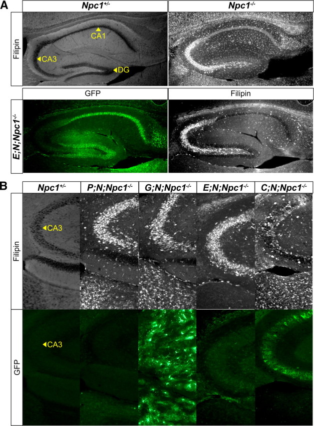
Neuron-autonomous correction of the CNS cholesterol storage defect in Npc1−/− mice. Immunofluorescence with anti-GFP marks neuron or astrocyte regions of the hippocampus that are positive for NPC1-YFP. Filipin marks hippocampus areas that are positive for accumulated free cholesterol. A, Sagittal sections of the hippocampus from an adult Npc1−/− mouse had substantial filipin staining in the CA3 neuron region. In an E; N; Npc1−/− mouse, NPC1-YFP produced in the DG or CA1 neurons did not reduce cholesterol accumulation in CA3 neurons, where NPC1-YFP expression was weak or absent. B, NPC1-YFP present in CA3 neurons (bottom) reduced CA3 cholesterol accumulation (top) in a C; N; Npc1−/− mouse. Despite NPC1-YFP production throughout the hippocampus in a G; N; Npc1−/− mouse, cholesterol accumulation was not reduced in CA3 neurons or elsewhere in the surrounding area.