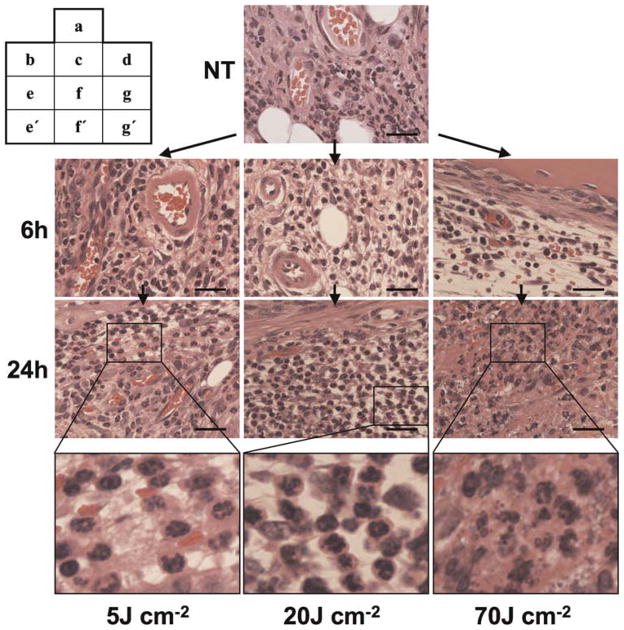
Fig. 6.
Histopathological images of knee joints of murine MRSA arthritis models. The lesions were collected without Photofrin® administration or photoirradiation (NT group, a), 6 hours after photoirradiation (b–d) and 24 hours after photoirradiation (e–g, e′, f′, g′), HE staining. Before photoirradiation (a), many polymorphonuclear leukocytes (neutrophils) with normal appearance are seen in proliferated synovial tissue. No morphological change or variation in number of neutrophils is seen in the PDT-treated knee joints with a fluence of 5 J cm−2 at each time (b, e, e′), whereas no morphological change are seen in the PDT-treated knee joints with a fluence of 20 J cm−2 at 6 hours after photoirradiation (c), and a remarkable increase of morphologically intact neutrophils is seen 24 hours after photoirradiation (f, f′). However, in the case of PDT with a fluence of 70 J cm−2, blood vessel destruction complicated with hemorrhage is seen at 6 hours after irradiation (d), and nonspheroidal leukocytes and atrophic changes of the synovium are seen at 24 hours after irradiation (g, g′). Bar = 30 μm.

