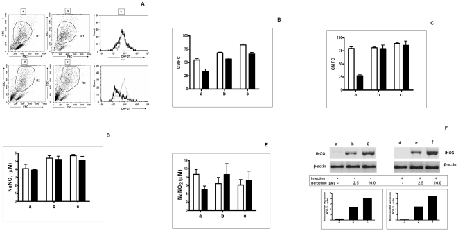
Figure 2. Effect of Berberine chloride on generation of NO and expression of iNOS.
A: A representative dot plot of uninfected (a) and Leishmania infected (d) murine peritoneal macrophages, that were treated with Berberine chloride (10 µM, 48 h, b, e). Cells were gated on the basis of characteristic linear forward and side scatter features of macrophages and subsequently DAF-2T fluorescence was measured on a logarithmic scale in the FL1 channel. A representative histogram of uninfected macrophages (c,  ) and L. donovani infected macrophages (f,
  ) for DAF-2T that were treated with Berberine chloride (…) macrophages as described in Methods. B: Uninfected macrophages (1×10⁶/ml, □, a) or L. donovani infected macrophages (▪, a) were treated for 24 h with Berberine chloride 2.5 µM (b) and 10 µM (c), and processed for measurement of DAF-2T fluorescence as described in Methods. Data are expressed as the mean GMFC ± SEM of at least 3 experiments in duplicate. C: Uninfected macrophages (1×10⁶/ml, □, a) or L. donovani infected macrophages (▪, a) were treated for 48 h with Berberine chloride 2.5 µM (b) and 10 µM (c) and processed for measurement of DAF-2T fluorescence as described in Methods. Data are expressed as the mean GMFC ± SEM of at least 3 experiments in duplicate. D: Uninfected macrophages (1×10⁶/ml, □, a) or L. donovani infected macrophages (▪, a) were treated for 24 h with Berberine chloride 2.5 µM (b) and 10 µM (c) and assayed for levels of extracellular NO as described in Methods. Each point represents the mean ± SEM of NO2
− (µM) of at least 3 experiments in duplicate. E: Uninfected macrophages (1×10⁶/ml, □, a) or L. donovani infected macrophages (▪, a) were treated for 48 h with Berberine chloride 2.5 µM (b) and 10 µM (c) and assayed for levels of extracellular NO as described in Methods. Each point represents the mean ± SEM of NO2
− (µM) of at least 3 experiments in duplicate. F: Uninfected macrophages (a) and L. donovani infected macrophages (d) were treated for 18 h with Berberine chloride 2.5 µM (b, e) or 10 µM (c, f). RNA was isolated and subjected to RT-PCR and the products of β-actin and iNOS mRNA were resolved on an agarose gel (1.5%) and quantified densitometrically using Total lab software as described in Methods.

