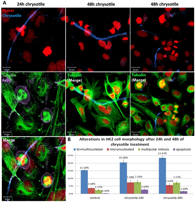
Figure 3. Alterations in the morphology of HK2 cells after 24 h or 48 h of chrysotile treatment.
Cells were processed by immunofluorescence to visualize nuclei, actin filaments and microtubules, and chrysotile fibers were observed by their autofluorescence. A) Confocal images of HK2 cells treated with chrysotile for 24 h or 48 h showing long, thick fibers interacting with the cells and multipolar mitosis; B) the alterations in cell morphology were analyzed, and after 24 h or 48 h of chrysotile treatment, the number of binucleated and multinucleated cells increased, as well the number of micronucleated cells, apoptotic cells and cells in multipolar mitosis (P<0.001).

