
Figure 6.
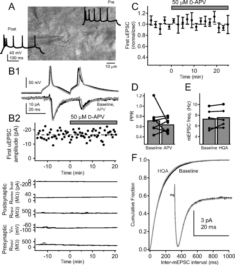
Functional PreNMDARs are absent from local synapses between L4 excitatory cells. A, Differential interference contrast image of example synaptically coupled L4 excitatory cells, with regular-spiking pattern for these cells. B1, Postsynaptic uEPSCs elicited by a pair of presynaptic spikes before (black) and after (gray) 50 μm d-APV application for the regular-spiking pair above. Each trace shown is the average of the last 10 sweeps of each condition. B2, Top, Lack of effect of d-APV on amplitude of the first uEPSC for one representative cell pair. Bottom: Postsynaptic holding current, postsynaptic series resistance, postsynaptic input resistance, presynaptic membrane potential, and presynaptic input resistance for this pair. C, Mean effect of d-APV application on first uEPSC amplitude (n = 7 pairs). D, Effect of d-APV on PPR. Error bars show population means. E, Mean mEPSC frequency for five L4 cells before and after 20 μm HQA application. F, Cumulative probability histogram of mEPSC interval before (black) and after (gray) HQA application (p > 0.1, Kolmogorov–Smirnov test). Inset, Mean mEPSC before (black) and during (gray) HQA application.