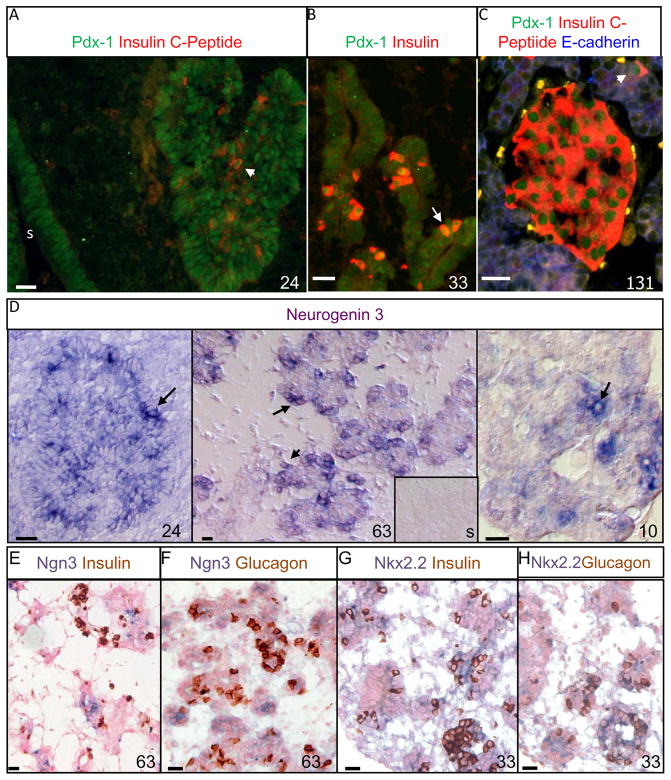
Figure1. Developmental Progression of Transcriptional Regulatory Factors in Sheep.
Immunofluorescent staining for Pdx-1 (Cy2, green) and insulin (Texas Red) was determined in the sheep pancreas at several stages of gestation (A–C). A longitudinal cross section at 24 dGA of the dorsal pancreatic bud and stomach (s) are shown (A), and the arrow identifies a Pdx1+ insulin+ cell. At 33 dGA elongated ducts composed of Pdx1+ epithelium were surrounded by a mesenchymal bed (B), solitary insulin+ cells with nuclear Pdx1 staining are apparent whereas insulin− cells exhibit a variety of Pdx1 staining localized in the nucleus and/or cytoplasm. Both mature islet-like structures and solitary insulin+ cells (arrow) are shown at 131 dGA (C), and E-cadherin staining in blue is observed in the epithelium. Representative photographs of sheep pancreas with differential interference contrast microscopy are shown for in situ hybridization with antisense or sense (insert) DIG-labeled neurogenin3 RNA for fetuses at 24 dGA and 63 dGA, and a lamb at postnatal day 10 (D). The black arrows identify examples of ngn3+ cells stained with BM purple. The inserted picture illustrates the negative control, Ngn3 sense strand (s), for the 63 dGA fetus and encompasses an area equivalent to the area of the antisense picture. A 63 dGA sheep pancreatic section were stained for Ngn3 by in situ hybridization techniques and subsequently immunostained for insulin (E) or glucagon (F). Pancreas section from a sheep fetus at 33 dGA was stained for Nkx2.2 using in situ hybridization methodologies and then immunostained for insulin (G) or glucagon (H). In all photographs, the factors are labeled above in their respective color, the gestational or postnatal age is indicated in the right corner, and a 20 micron scale bar is present in the lower left corner.

