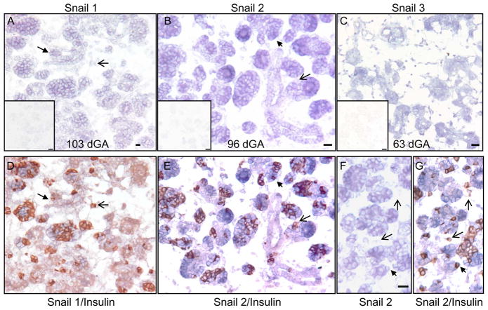
Figure 6. Expression of the Snail Family Members in the Sheep Pancreas.
On sheep pancreatic sections, in situ hybridizations were performed with antisense and sense (inserts) Snail 1 (A), Snail 2 (B & F), and Snail 3 (C) cRNA. After capturing images for Snail 1 and Snail 2, the sections were subsequently immunostained for insulin to co-localize Snail 1 (D) and Snail 2 (E & G) to β-cells. The black filled arrows identify β-cells co-expressing Snail 1 or Snail 2. The open arrowheads identify Snail-insulin+ cells. Scale bars in the antisense in situ hybridization pictures represent a 20 micron and bars in the inserts (sense strand, negative controls) are 50 microns long.

