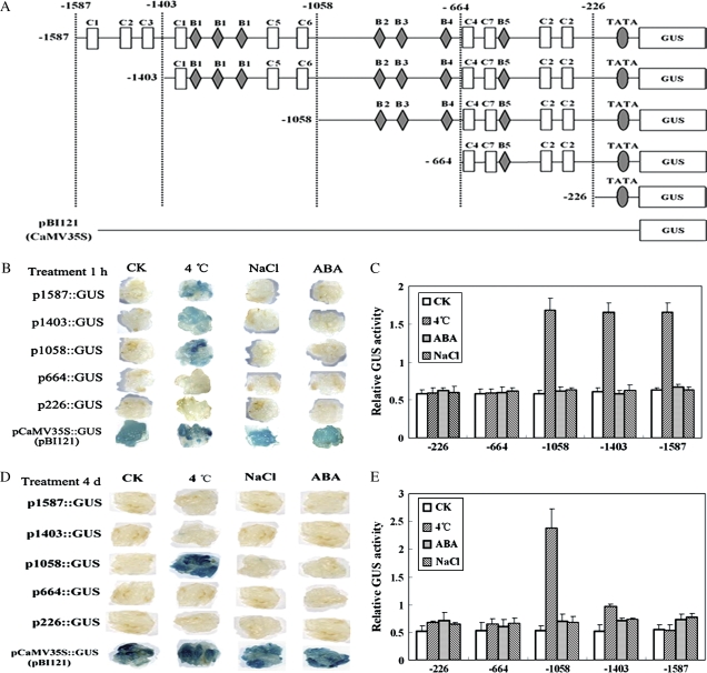
Fig. 2.
Activity analysis of the GmDREB3 promoter. (A) Five fragments that were deleted from the 5′ end of the GmDREB3 promoter were inserted into a binary vector (pBI121). The locations of the 5′ ends of the five fragments of the GmDREB3 promoter are indicated; ‘C’ and ‘B’ represent MYC and MYB recognition sites, respectively. Binary vector pBI121 was used as the positive control. (B and D) After transformation with vectors containing the five fragments of the GmDREB3 promoter, calli were treated on MS medium plus 100 μM ABA (ABA), plus 200 mM NaCl (NaCl), and under low temperature (4 °C) stress and normal conditions (CK) for 1 h and 4 d. The histochemical staining results (GUS) are shown. (C and E) Quantitative results of GUS activity after treatments for 1 h and 4 d. The relative GUS activity was calculated as the ratio of GUS activity of a mutant GmDREB3 promoter to that of the CaMV 35S promoter (pBI121) under the same stress treatments.

