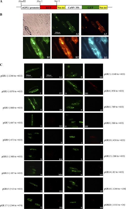
Fig. 5.
(A) The schematic map of RFP/GFP double marker genes in the transient expression vector pBI221 with CaMV 35S promoter-driven GFP and RFP. (B) CaMV 35S promoter-driven GFP and RFP transient expression in onion epidermal cells. The results shown are from transient transformed epidermal cells using the DNA delivery particle bombardment system. (A) Light images of transiently transformed epidermal cells. (A1) Fluorescent images of (A) under an exciter filter (450–490 nm) that expressed GFP. (A2). Fluorescent images of (A1) under an exciter filter (510–560 nm) that expressed RFP. (B1) Fluorescent images that expressed GFP. (B2) Fluorescent images of (B1) that expressed RFP. (B3) Fluorescent images of (B1) under an exciter filter (380–420 nm). (C) Variable-length truncated AGPL1 promoter-driven RFP transient expression in epidermal cells. Single letters: fluorescent images expressed GFP driven by the CaMV 35S promoter. Double letters: fluorescent images expressed RFP driven by the AGPL1 promoter deletions. (This figure is available in colour at JXB online.)

