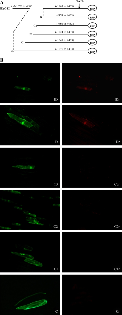
Fig. 6.
(A) A schematic map of RFP/GFP double marker gene transient expression vectors harbouring different fine truncated deletions of the AGPL1 promoter. ID (C and D), pGRID (–1140 to +433, –1070 to –959 deleted); C, pGR3 (–1070 to +433); C1, pGR31 (–1047 to +433); C2, pGR32 (–1024 to +433); C3, pGR33 (–986 to +433); D, pGR4 (–958 to +433). (B) C–ID are fluorescent images under an exciter filter (450–490 nm). Cr–IDr are fluorescent images of (C–ID) under an exciter filter (510–560 nm). All images were observed under 40× magnification. (This figure is available in colour at JXB online.)

