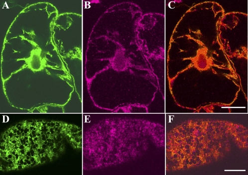
Fig. 5.
Localization of GFP-labelled ER and the 175 kDa myosin in interphase cells. Immunofluorescence staining with the antibody against 175 kDa myosin was carried out for cells expressing GFP-labelled ER. (A and D) GFP–ER. (B and E) 175 kDa myosin. A, B, and C, and D, E, and F, were focused at nuclear and cortical regions, respectively. C and F are merged images of A and B, and D and E, respectively. The bar represents 20 μm.

