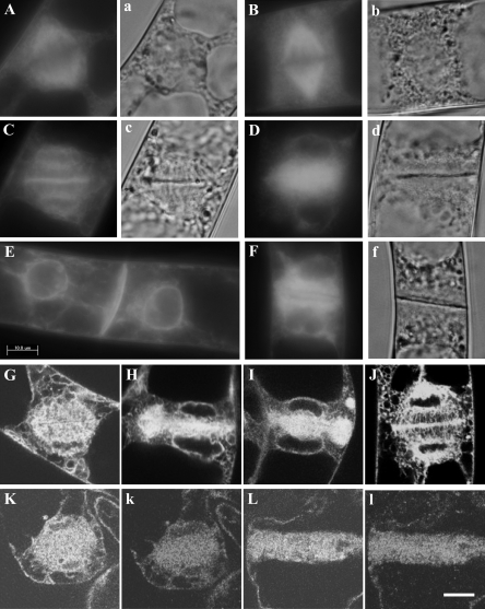
Fig. 9.
Suppression of ER accumulation in the equatorial plane and around daughter nuclei, and of positioning of daughter nuclei in the centre of daughter cells by BDM, and localization of 175 kDa myosin in BDM-treated cells. GFP–ER (upper case letters), differential interference contrast images (a, b, c, and f), and signals of 175 kDa myosin (k and l) are shown. After releasing cells from propyzamide, they were treated (B, D, F, G, H, I, K, L, k, and l) or not with 50 mM BDM (A, C, E, and J). The accumulation of GFP–ER in the spindle region was not suppressed by treatment with BDM (B), perhaps because spindle formation began soon after release from propyzamide. In mid telophase, GFP–ER accumulated in the equatorial plane of phragmoplast and around daughter nuclei in cells without BDM treatment (C and J). The cell plate was assembled between daughter nuclei (c). However, accumulation of GFP–ER in the equatorial planes and around daughter nuclei was suppressed by the treatment with BDM (D), although the cell plate was assembled (d). The suppression of those events was confirmed by observation using a laser scanning microscope (G, H and I). After mitosis of control cells, daughter nuclei moved to the centre of each daughter cell (E), whereas they did not in BDM-treated cells (F). The 175 kDa myosin (k and l) in the cells treated with BDM was in a similar localization pattern to GFP–ER (K and L). Bars represent 10 μm.

