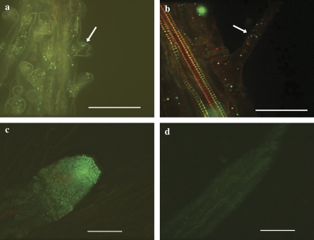
Fig. 5.
Localization of MT2 protein (arrows) in root epidermal cells and root hairs in (a) 7-d-old T. caerulescens; (b) 4-d-old Arabidopsis. The root tip of 4-d-old Arabidopsis (c) transformed with TcMT2 under the 35S promoter shows a bright fluorescence compared with (d) the wild type (Col-0). Immunocytochemical analysis was performed using primary antibodies raised against TcMT2. The secondary antibody was conjugated with Alexa Fluor 488, and fluorescent staining was imaged by laser-scanning confocal microscopy. The samples were stained with propidium iodide. Bar = 100 μM (c, d) or 50 μM (a, b). (This figure is available in colour at JXB online.)

