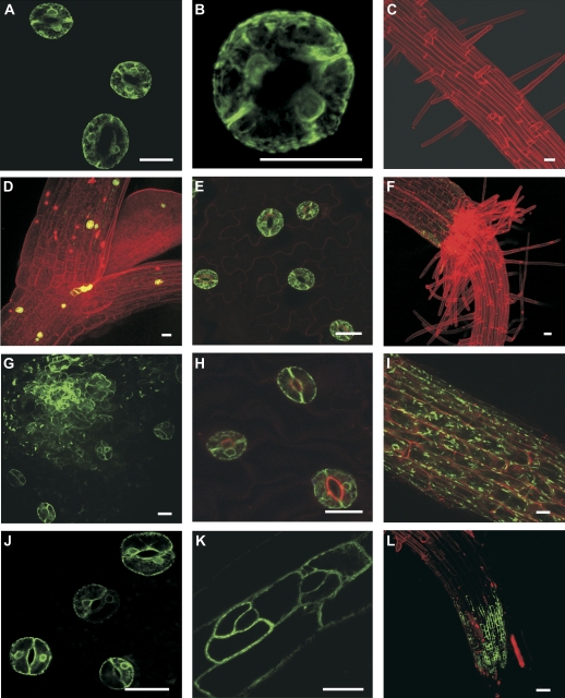
Fig. 2.
3-D projections of CLSM images of single insert enhancer trap lines stably expressing GFP in stomatal guard cells. E1728 (A–C) and E361-1 (D–F) had guard cell-specific GFP expression. Lines KS019-1 (G–I) and J2103-1 (J–L) had predominant guard cell GFP expression, but GFP was also detected in leaf and root epidermal cells. All scale bars represent 20 μm.

