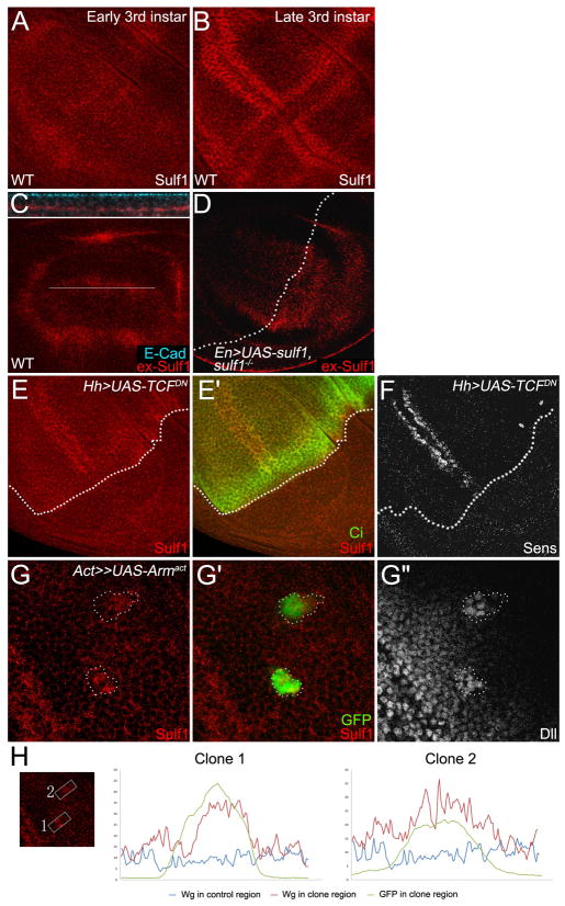
Fig. 1. sulf1 is dynamically expressed and its expression is up-regulated by Wg signaling.
A,B: Immuno-staining of 6-O endosulfase in wild-type wing discs. A: In early 3rd instar discs, sulf1 is ubiquitously expressed in entire wing disc. B: In late 3rd instar discs, expression of sulf1 is highly up-regulated in each side of DV compartment boundary and AP compartment boundary. C,D: Extracellular Sulf1 staining in wild-type wing disc (C) and in wing discs overexpressing sulf1 in the posterior compartment driven by En-Gal4 on sulf1 mutant background (D). The upper panel in C shows z-section of the line in lower panel. Staining of E-cadherin marks the apical side of the cells. High levels of extracellular Sulf1 are detected at basal-lateral regions. Dotted line in D shows AP compartment boundary. E: Sulf1 staining is eliminated in TCFDN expression domain marked by the absence of Ci (E′). F: Sens staining shows the loss of Wg signaling in TCFDN expression domain. AP boundary is outlined by broken lines. G: A gain-of-function Armadillo (Arm) Armact was ectopically expressed by using FLP-OUT system. Sulf1 staining is elevated in the clones (G) that are marked by GFP (G′). Dll staining shows enhanced Wg signaling activity in the clones (G″). The clones are outlined by broken lines. H: Signal profiles of Sulf1 (red line) and GFP (green line) are plotted from selected areas in the box and non-clone control region (blue line). Wing discs in C, D, E, F, G, H were all obtained from late 3rd instar larva. In all images, anterior compartment faces left, and dorsal compartment faces up.

