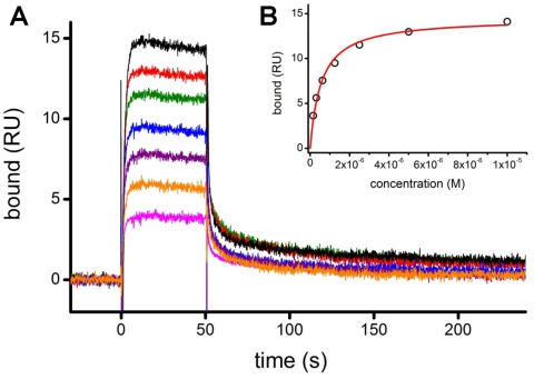
Figure 3. Surface plasmon resonance of rCR2.
(A) A representative SPR sensorgram of rCR2 binding to immobilized C3d. C3d was coupled to the SPR sensor chip using the free thiol of the Cys17 residue. Binding was determined over a range of rCR2 concentrations; shown here, 78 (pink), 156 (orange), 313 (purple), 625 (blue), 1250 (green), 2500 (green), 5000 (red) and 10000 nM (black). (B) The binding affinity of rCR2 to C3d was determined by plotting the binding response at equilibrium against the concentration of rCR2 and fitting a Langmuir binding isotherm.

