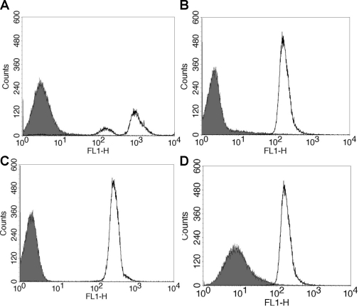
Figure 5. Flow Cytometry of rCR2 or anti-C3d bound to C3d+ red blood cells.
Sensitised sheep red blood cells were incubated with human C6 deficient serum to generate C3d deposition on RBC in the absence of cell lysis. (A) To confirm C3d deposition, C3d+ RBC incubated with anti-human C3d (black line) or isotype control (solid grey) followed by mouse anti-human-FITC secondary. (B) To demonstrate rCR2 binding to C3d, C3d+ RBC cells were incubated with rCR2 (black line) or an irrelevant His-tag protein, C2A, (solid grey) followed by secondary anti-HIS-FITC. (C) To further demonstrate that the mutant K41E CR2 mutant did not bind to C3d, C3d+ RBC cells were incubated with rCR2 (black line) or K41E CR2 (solid grey) followed by secondary anti-HIS-FITC. (D) To demonstrate that rCR2 specifically bound to C3d, C3d+ RBC cells were first incubated with the inhibitory anti-human C3d prior to incubation with rCR2 and secondary anti-HIS-FITC (solid grey) or as positive control rCR2 and secondary anti-HIS-FITC alone (black line).

