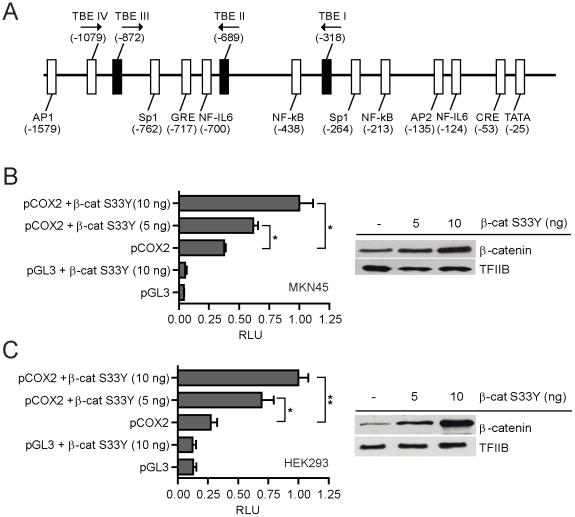
Figure 3. Human pCOX2-1.6 promoter activity in response to Wnt/β-catenin signaling.
(A) Schematic drawing depicting the genomic context of ca. 1.6 Kbp from the transcriptional start site (TSS) of the human COX2 promoter, including the location of known transcriptional regulators (white boxes) and the position of the three novel TCF/LEF-binding elements (TBE: core CTTTG; black boxes) determined in silico. (B & C) Gene reporter assays in MKN45 (B) and HEK293 (C) cells co-transfected with 10 ng of pCOX2 and increasing concentrations of a constitutively active β-catenin (S33Y) protein (left panel). Cells were co-transfected with 1 ng of PRL-SV40 Renilla as an internal control. Promoter activity was normalized as the ratio between firefly luciferase and Renilla luciferase units. RLU: Relative Luciferase Units. Each figure corresponds to at least three independent experiments. Statistical significance was determined through ANOVA test (* p<0.05, ** p<0.01). Nuclear levels of β-catenin protein were examined in same cell lines through Western Blot analysis (right panel). The TFIIB general transcription factor was used as an internal control.

