
Figure 3. Effects of gal-4 expression.
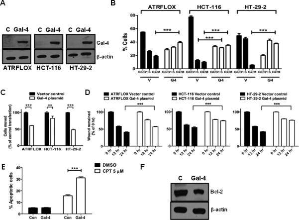
(A). Gal-4 transient transfection in ATRFLOX, HCT-116 and HT-29-2 cells. Cells were transiently transfected with either the gal-4 plasmid or the vector. Cell lysates (10 μg each) prepared after 24 h were analyzed for gal-4 and β-actin. (B). Cell cycle analysis. The above transiently transfected cells were also subjected to cell cycle analysis as described under Materials and Methods. The G1, G2/M and S phase distribution of cells was determined by FlowJo software. *** indicates P <0.001. (C). Cell motility assay. The above transiently transfected cells were used in motility assay as described under Materials and Methods. A P value of <0.001 was considered significant. ** indicates P <0.01 and *** indicates P <0.001. (D). Wound healing assay. The above transiently transfected cells were used in migration assay as described under Materials and Methods. A P value of <0.001 was considered significant. *** indicates P <0.001. (E). Effect of gal-4 on CPT induced apoptosis. HCT-116 cells transfected with either the vector plasmid or the gal-4 plasmid for 48 h and were subjected to apoptosis in the presence of either DMSO (vehicle control) or CPT (5 μM) for 4 h. *** indicates P <0.001. (F). Effect of gal-4 on Bcl-2 expression. HCT-116 cells were transiently transfected with either the vector plasmid or the gal-4 plasmid and the expression of Bcl-2 was analyzed by western blotting after 48 h. β-actin was used as loading control.