
Figure 6. Expression analysis of β-catenin in normal and adenocarcinoma sections.
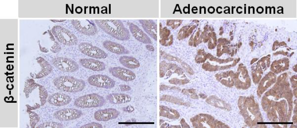
Immunohistochemistry was carried out on human colon normal and adenocarcinoma sections and β-catenin staining was visualized by DAB (brown). Magnification was 20x and the scale bar represents 500 μm.