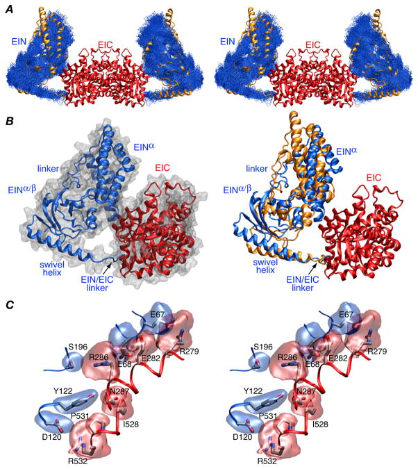
Figure 2.
Structure of the EI(H189Q) dimer determined from RDC and SAXS/WAXS data. (A) Stereoview of a best-fit superposition (to the EIC dimerization domain which remains fixed) of the final 100 simulated annealing structures calculated with random noise added to the RDCs with the backbone (N, Cα, C′) atoms of the EIN domain in blue, the EIC domain displayed as a red ribbon, and the position of the EIN domain in wild-type EI9 displayed as a gold ribbon. (B) Ribbon diagram of a single subunit of EI(H189Q) with the EIN and EIC domains in red and blue, respectively, a molecular surface shown in grey in the left panel and the position of the EIN domain in wild-type EI in gold in the right panel. The y axis of the RDC alignment tensor coincides with the C2 symmetry axis of the dimer. (C) Stereoview of the side chain interactions at the EIN/EIC interface in the EI(H189Q) mutant with reweighted atomic probability maps20 for the side chains of EIN and EIC shown in blue and red, respectively (plotted at a threshold of 15% of maximum and calculated from the 100 final simulated annealing structures).

