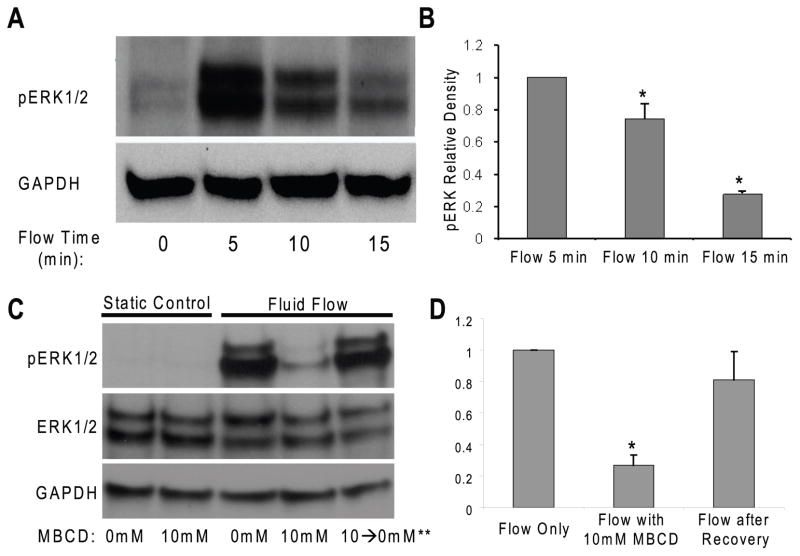
Figure 2. MβCD inhibits oscillatory fluid flow induced ERK1/2 phosphorylation in MC3T3-E1 osteoblastic cells.
A, a dramatic increase in ERK1/2 phosphorylation was observed at 5, 10 and 15 minutes in response to oscillatory fluid flow (1N/m2, 1Hz). B, bar graph representation of ERK1/2 phosphorylation quantified by scanning densitometry normalized to GAPDH. C, after 10mM MβCD treatment, oscillatory fluid flow induced phosphorylation of ERK1/2 at 5 minutes significantly decreased in MC3T3-E1 osteoblastic cells (lane 1–4). Replacement of MβCD media with control flow media for 120 minutes allowed the recovery of cell responsiveness to fluid flow in terms of ERK1/2 activation (lane 5). D, bar graph representation of ERK1/2 phosphorylation quantified by scanning densitometry normalized to GAPDH. (*, p<0.05) Each bar represents the mean ± S.E. and each experiment was repeated on 3–5 times.

