
Figure 2.
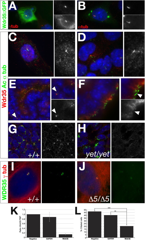
Wdr35 Localizes to Cilia and Is Required for Ciliogenesis
(A and B) NIH-3T3 (A) or IMCD3 (B) cells were microporated with full-length mouse Wdr35::GFP and serum starved for 36 hr before costaining with antibodies directed against γ-tubulin (A) or acetylated α-tubulin (red; B). Nuclei are stained with DAPI (blue). Magnification of regions of interest are shown in singl-channel images indicating colocalization.
(C and D) IMCD3 cells were serum starved for 36 hr before costaining with antibodies directed against acetylated α-tubulin (green) and Wdr35 (red; C, 085 antibody; D, 02 antibody). Nuclei are stained with DAPI (blue).
(E and F) Wild-type 13.5 dpc mouse kidney (E) and choroid plexus (F) sections were costained with antibodies directed against acetylated α-tubulin (green) and Wdr35 (red, 085 antibody). Nuclei are stained with TOTO-3 (blue). Primary cilia in the mesenchyme (E) and ciliated epithelia lining lumen (F) are indicated.
(G and H) Shown are 0.4 μm confocal images of section immunohistochemistry of 11.5 dpc limb-bud mesenchyme from wild-type (G) and Wdr35yet/yet (H) embryos stained with antibodies directed against acetylated α-tubulin (green), Wdr35 (085 antibody: red), and TOTO-3 (blue). See also Figure S3E for additional support, by immunoblot analysis, that yeti is a null allele of Wdr35. See also Figure 4 for preadsorbtion with peptide for demonstration of the specificity of antibody studies.
(I and J) Primary fibroblast cells from a control (I) or WDR35Δ5/Δ5 SRP patient (J) were serum starved for 36–48 hr before costaining with antibodies directed against WDR35 (green, 02 antibody) and γ-tubulin (red). Fainter, nonspecific staining of cytoplasmic microtubules by γ-tubulin is observed in human control and mutant fibroblasts. Nuclei are stained with DAPI (blue).
(K and L) Wdr35 siRNA knockdown leads to reduced cilia formation. ShhLIGHT II cells were transfected with siRNAs against Wdr35. qRT-PCR shows significantly reduced levels of Wdr35 mRNA after siRNA treatment (K). A 50% reduction in the number of ciliated cells was observed when Wdr35 mRNA was knocked down to 15% of wild-type levels (L). Negative: scramble siRNA; ∗∗∗p < 0.001; ∗∗p < 0.01 (Chi-squared). Bars represent standard deviations. Statistical significance was determined via a Student's t test.