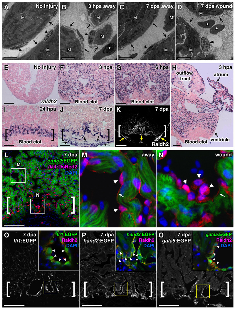
Figure 1. Resection of the Ventricular Apex Stimulates Immediate, Organ-wide Morphological and Molecular Changes in Endocardium.
(A–D) Transmission electron microscope (TEM) analyses of endocardium in uninjured (A) and injured ventricles (B–D). Arrowheads, endocardial nuclei. Arrows, endocardial cell bodies. M, cardiac muscle. Asterisk, red blood cell. Scale bar, 2 µm.
(E–K) raldh2 expression assessed by in situ hybridization (E–J) and Raldh2 immunostaining (K) in uninjured (E) and injured (F–K) ventricles. Brackets in (I–K), injury site. Arrows in (J, K), epicardial cells. Scale bar (E–Q), 100 µm.
(L–N) Confocal images of altered endocardial cell shape, and enhanced flk1 driven DsRed2 fluorescence in the injury site (A, brackets) in a 7 dpa cmlc2:EGFP; flk1:DsRed2 double transgenic ventricle. Arrowheads, endocardial nuclei. Arrows, endocardial lining of myofiber. An antibody against DsRed was used. DAPI (4'-6-Diamidino-2-phenylindole) stains nuclei.
(O–Q) Sections of 7 dpa fli1:EGFP (O), hand2:EGFP (P), or gata5:EGFP (Q) transgenic ventricles. Brackets, injury sites. Arrowheads in inset, Raldh2+/EGFP+ endocardial nuclei with rounded morphology (G–I).

