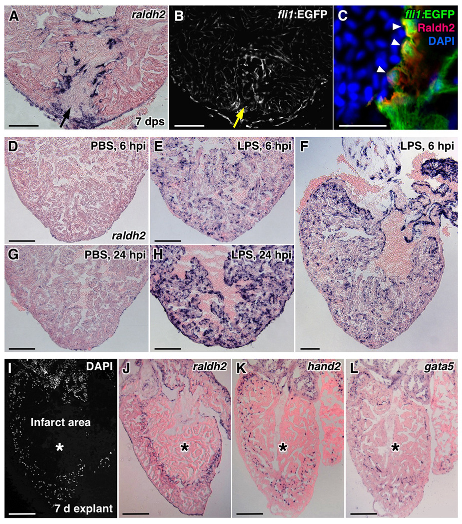
Figure 2. Induction of raldh2 Expression in Various Injury Models.
(A,B) Stab injuries into the ventricular apex assessed for raldh2 induction (A) and fli1:EGFP expression (B) at 7 days post-stab (dps). Arrows, needle entry site. Scale bar, 100 µm.
(C) Confocal image of Raldh2 immunofluorescence in fli1:EGFP+ endocardial cells with rounded morphology at the injury site (arrowheads). Scale bar, 20 µm.
(D–H) raldh2 induction after intraperitoneal LPS or vehicle (PBS) injection. Scale bar, 100 µm (D–L).
(I–L) Endocardial raldh2 (J), hand2 (K), and gata5 (L) expression surrounding spontaneous infarcts (asterisks) within cultured ventricular explants. Dead cardiac tissue was identifiable by the absence of cell nuclei (I).

