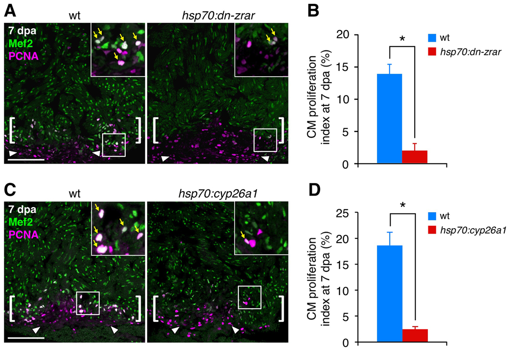
Figure 3. Transgenic Inhibition of RA Signaling Blocks Cardiomyocyte Proliferation during Regeneration.
(A) Assessment of Mef2+PCNA+ cells (arrows) in wild-type (wt) and hsp70:dn-zrar transgenic fish at 7 dpa, after a single heat-shock at 6 dpa. Maximum projection images of 10 µm z-stacks are shown. Insets, high-magnification images of the rectangle. Arrowheads, proliferating epicardial cells. Brackets, injury site. Scale bar, 100 µm (A, C).
(B) Quantification of CM proliferation in wt and hsp70:dn-zrar transgenic fish at 7 dpa. Data are mean ± SEM from 6 animals each (3097 wt and 2482 transgenic CMs analyzed). *p < 3 × 10⁻⁵, Student's t-test.
(C) Assessment of Mef2/PCNA double-positive cells (arrows) in wt and hsp70:cyp26a1 transgenic fish at 7 dpa, after a single heat-shock at 6 dpa. Maximum projection images of 10 µm z-stacks are shown.
(D) Quantification of CM proliferation in wt and hsp70:cyp26a1 transgenic fish at 7 dpa. Data are mean ± SEM from 4 wt and 6 transgenic animals (3888 wt and 4760 transgenic CMs analyzed). *p < 2 × 10⁻⁴, Student's t-test.

