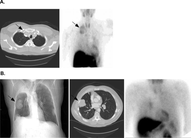
Figure 3.
99mTc-Sestamibi scans performed in two patients with NSCLC. A: CT scan (left image) shows a large supraclavicular mass (denoted by arrow) that also exhibits 99mTc-Sestamibi uptake (arrow, left image). B: Chest X-ray (left image) and CT scan (center image) clearly show a peripheral right lung lesion (denoted by arrow) that does not uptake 99mTc-Sestamibi (right image).

