
Figure 1. RIP1 deficiency rescues FADD−/− mice from embryonic necrosis and lethality.
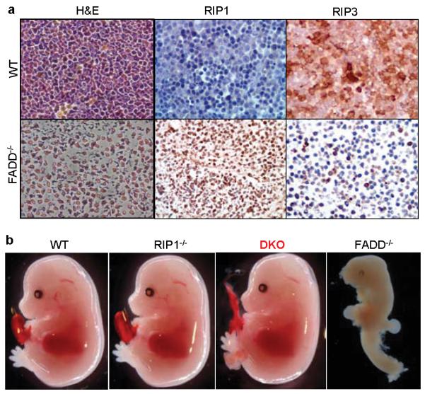
a, FADD−/− embryos exhibit massive necrosis and altered RIP1 and RIP3 expression. E12.5 wild type (WT, top panels) or FADD−/− embryos (bottom panels) were fixed in formalin. Left panels of hematoxylin and eosin (H&E) staining show extensive cell loss and pyknotic nuclei in the FADD−/− fetal liver. Middle and right panels indicate staining for RIP1or RIP3. b, E14.5 DKO embryos appear normal, contrasting the defective FADD−/− embryos.