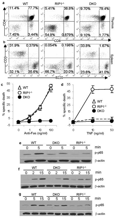
Figure 2. FADD deficiency partially corrects the RIP1−/− T cell developmental defect by blocking apoptosis.
Lymphocytes (a-b) in the chimeras of the indicated genotypes were analyzed. E18.5 fetal thymocytes of the indicated genotypes were treated with anti-Fas antibodies (c) or TNFα (d) and death responses determined 12 hours post stimulation. Error bars represent mean ± SEM of triplicates. T cells (e) and B cells (f) from NSG chimeras of the indicated genotypes were stimulated with anti-CD3 and anti-CD28 antibodies or with LPS, respectively. MEFs were stimulated with TNFα (g). NF-κB activation was indicated by the induction of p65 phosphorylation. β-actin, loading controls.

