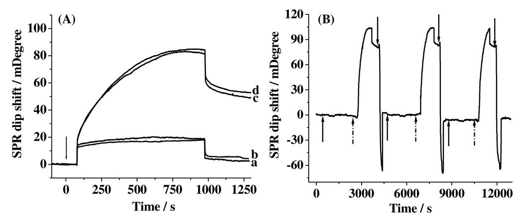
Figure 3.
SPR sensorgrams after injections of 30.00 nM detection conjugate to Aβ(1–40) capture antibody (a) before and (b) after exposed to 0.50 nM Aβ(1–42), (c) Aβ(1–42) capture antibody that had been exposed to 0.20 nM Aβ(1–42), and (d) same as (a) but with exposure to 0.20 nM Aβ(1–40). The arrow indicates the time when the injections were made. (B) A sensorgram showing three repeated cycles for the injections of 0.50 nM Aβ(1–40) (signified by the solid upward arrows) and 30.00 nM detection conjugate (starting from the dotted arrows) into an SPR channel covered with the Aβ(1–40) capture antibody and the regeneration of the sensor surface using 10 mM NaOH (starting from the downward arrows).

