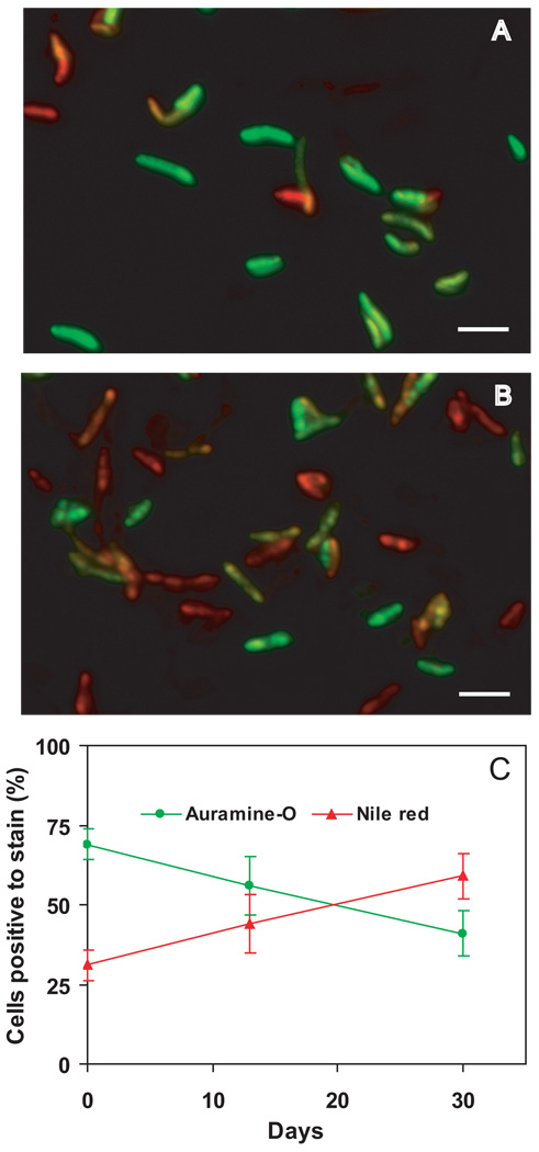
Fig. 6. Accumulation of lipid bodies and loss of acid-fastness in M. tuberculosis cells during hypoxia.
M. tuberculosis cultures were subjected to gradual O2 depletion (Wayne model). Aliquots from mid-log cultures (panel A) and cultures at day 30 of the hypoxic time course (anaerobiosis) (panel B) were stained for acid-fastness with Auramine-O (green) and for lipid body accumulation with Nile Red (red). Cells were examined by fluorescence microscopy at the same intensity for all samples with Z stacking to get the depth of the scan field. Overlaid images of Auramine-O- and Nile-Red-stained cells are shown. Bar = 4µM. Panel C: Cells stained with Auramine-O (green line) or Nile-red (red line) were counted from three microscopic fields. Means (and SD) of % stain-positive cells are shown.

