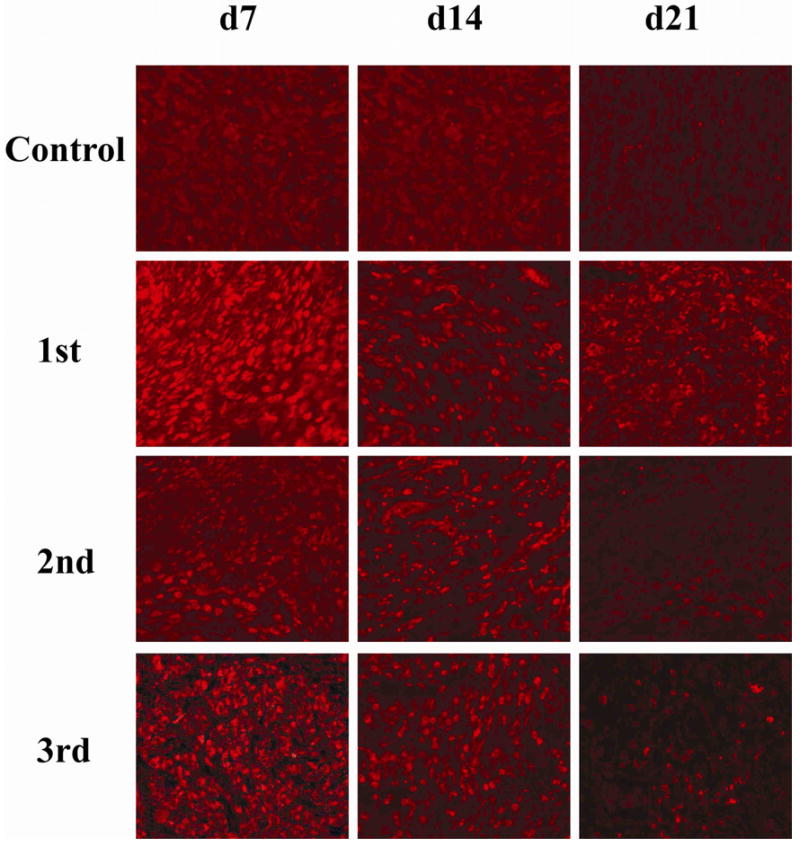
FIG. 1.

Adenoviral type 5 RIP-TK expression in PANC-1 tumors.
A-5-RIP-TK was intravenously administered to SCID mice 2 mo after inoculation with PANC-1 human pancreatic cancer cells. L-A-5-RIP-TK was repeated administrated on day 22, 43 after initial delivery, three gene deliveries in total. Mice were sacrificed on day 7, 14, 21d after each A or L-A-5-RIP-lacZ administration, respectively. The tumor tissues were processed and sectioned as usual. Immunostaining was performed using anti-HSV-TK antibody. Red staining cells indicate strong expression of HSV-TK in tumor cells. The figure showed the TK gene expression profile in control group (top row), first gene delivery (next to top row), second delivery (third row from top) and third delivery (bottom row) (x200)
