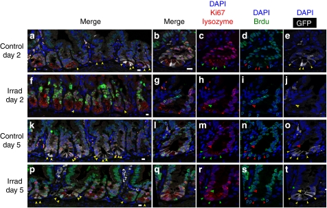
Figure 6. Immunofluorescence CBC cell BrdU-labelling at 2 and 5 days post irradiation.
Control (a–e,k–o) and irradiated (f–j,p–t) Lgr5-EGFP mice were continuously exposed to BrdU administered in drinking water and killed after 2 and 5 days. Ileum sections were simultaneously stained for nuclear BrdU (green), nuclear Ki67 (red), cytoplasmic lysozyme (red) and cytoplasmic green fluorescent protein (GFP, white). Nuclei were counterstained with DAPI (blue). Closed and open arrowheads point at CBC and to cp4 cells, respectively. (a) Merge picture of neighbouring GFP+ and GFP− crypt sections. (b) Merge picture of a crypt section with four CBC cells staining positive for Ki67 (c, green arrowheads) BrdU (d, red arrowheads) and GFP (e, yellow arrowheads). (f) Merge picture of crypt sections devoid of CBC cells. (g) Merge picture of a single crypt section. The cp4 cell of this crypt (open arrowhead) is Ki67+ (h), BrdU+ (i) and GFP− (j). (k) Merge picture of GFP+ crypt sections. (l) Merge picture of a single crypt section with four CBC cells and one cp4 cell, all of which are Ki67+ (m), BrdU+ (n) and GFP+ (o). (p) Merge picture of a succession of GFP+ and GFP−crypt sections. (q) Merge picture of a single GFP+ crypt section with four CBC cells, all of which are Ki67+ (r), BrdU+ (s) and GFP+ (t). Note the presence of a Ki67+ BrdU+ GFP− cp4 cell and the BrdU+Ki67− nucleus (*) of a post-mitotic cell sandwiched between Ki67+ BrdU+ progenitor nuclei. P, Paneth cell nuclei. Scale bar, 10 μm. DAPI, 4′-6-diamidino-2-phenylindole.

