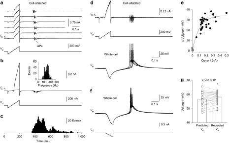
Figure 1. Ensemble potassium channel activity in cell-attached patches triggers action potential firing.
(a) Tiled consecutive traces of action potential (AP) firing triggered by ensemble potassium channel activity in a cell-attached (IC-A) patch made from the soma of a visually identified thalamocortical (TC) neuron. The lower trace shows the voltage command ramp (Vp). (b) Digital average (n=30 responses) of ensemble potassium channel activity evoked in the cell-attached patch shown in a. The fitted grey line represents the leak current. The inset shows the instantaneous AP firing frequency. (c) Post-stimulus time histogram of AP firing generated by ensemble potassium channel activity in cell-attached patches (data pooled from 20 neurons). (d) Simultaneous cell-attached (IC-A) and whole-cell current-clamp (Vm) recording from the soma of a TC neuron. Five consecutive traces have been overlain. (e) Relationship between the peak amplitude of ensemble potassium channel activity and intracellular voltage responses, for cell-attached pipettes filled with a potassium-based solution (black symbols) or a solution that included the potassium channel blockers 4-aminopyridine and tetraethylammonium (grey symbols). Data have been fit by an exponential function (line). (f) Voltage responses (Vm) evoked by the injection of a ramp of negative current through the recording electrode (Iinj) during somatic whole-cell current-clamp recording. Five consecutive traces have been overlain. (g) Resting membrane potential of TC neurons determined by whole-cell current-clamp recording (recorded) or predicted by measurement of the reversal potential of ensemble potassium channel activity in cell-attached patches (n=28). Data were compared with a paired Student's t-test.

