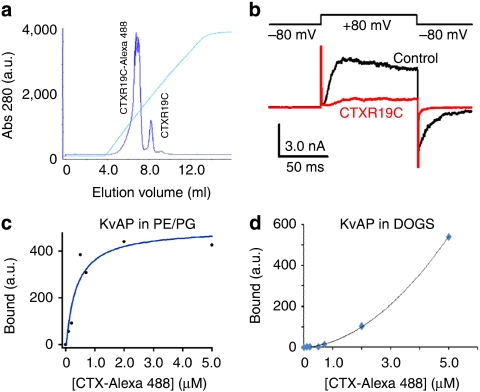
Figure 3. Channels in DOGS vesicles appear folded properly.
(a) Elution of CTX-R19C-Alexa 488 from a Resource S FPLC column. The ionic gradient (cyan trace) of NaCl varied from 30 to 500 mM. The conjugate eluted at ~150 mM NaCl with a noisy peak tip due to detector saturation. (b) Recombinant CTXR19C was active on the KvAP channels in lipid bilayers. In all, 200 nM CTXR19C was added to the extracellular side of the bilayer, which inhibited almost all channel activity after 10 min (red trace against the black control). (c) CTXR19C-Alexa 488 binding to the KvAP channels in PE/PG vesicles. Vesicles were incubated with CTX-Alexa 488 in a small volume, and then pelleted down at 250,000 g for 45 min. The pellet was resuspended in a DM buffer for fluorescence measurement. For each data point (black dots), a control sample with 100 μM CTX was prepared to measure nonspecific binding, and specific binding was plotted as a function of the CTX-Alexa 488 concentration. Fitting of the data (blue line) with a Michaelis–Menten equation yielded a kD of 350 nM, lower than unconjugated toxin. (d) Binding of CTX-Alexa 488 to the KvAP channels in DOGS vesicles (blue diamonds with a black line from polynomial fitting). Even though the DOGS membranes decrease the affinity of KvAP to CTX-Alexa 488 (kD > 2.0 μM), there was still high specific CTX-binding at 2.0 and 5.0 μM of CTX-Alexa 488.

