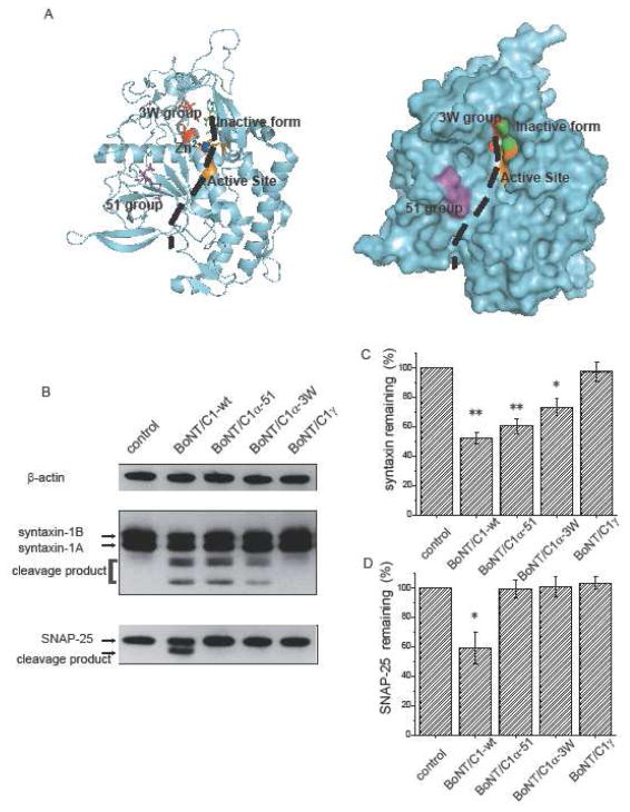
Fig. 1.
A. Structure of BoNT/C1 light chain (12) in ribbon (left) and space-filling (right) representations (PDB ID: 2QN0). The position of the substrate, indicated by a black dashed curve, was inferred from the BoNT/A-substrate crystal structure (11). B. Western blot analysis of extracts from cultured hippocampal neurons blotted for syntaxin and SNAP-25. Untransfected control cells (lane 1) show full length syn-taxin 1A, syntaxin 1B, and SNAP-25. Infection with wild type BoNT/C1 light chain cleaves all three proteins (lane 2). BoNT/C1α-51 (lane 3) and BoNT/C1α-3W (lane 4) cleaved syntaxin and spared SNAP-25. BoNT/C1γ showed no catalytic activity toward any substrate. C and D. Gel quantification of BoNT/C1 cleavage. The syntaxin (C) and SNAP-25 (D) band densities were reduced by cleavage in parallel with the appearance of lower molecular mass cleavage products. Syntaxin and SNAP-25 signals were normalized to the β-actin control and averaged (N=3).

