
Fig. 7.
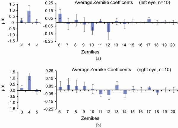
Mean values of 2nd to 5th order Zernike modes for 20 eyes across a 2 mm pupil, using wavefront spots focused on the outer retina. Zernikes for the 10 left eyes are shown in (a) and ones for the 10 right eyes are shown in (b). Error bars represent ± 2 times SEM. Lower and higher order aberrations are shown using different scales.