
Fig. 12.
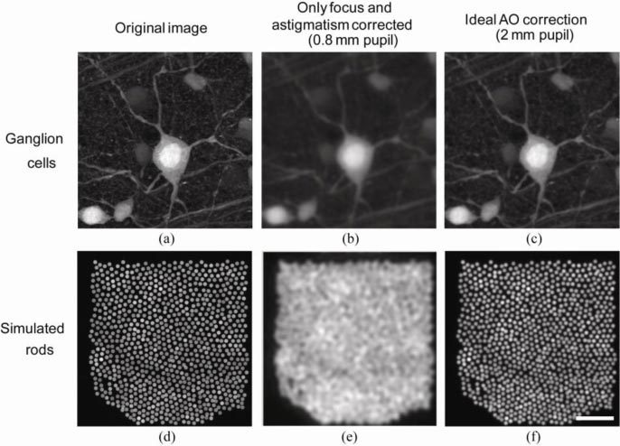
Simulated retinal images of ganglion cells and rod photoreceptors under ideal imaging conditions and in the absence of noise with/without AO correction. (a) A fluorescent ganglion cell image from a flat-mounted rat retina imaged with confocal microscopy with 1.2 NA. (b) A simulation of the retina in (a) being imaged without AO correction over a 0.8 mm pupil under the best possible condition. (c) A simulation of the same ganglion cell being imaged with AO correction over a 2 mm pupil under the best possible condition. (d) A simulated rod photoreceptor mosaic in the mouse retina. (e) & (f) are the same without AO and with AO simulations of the rod photoreceptor mosaic. Scale bar: 20 µm.